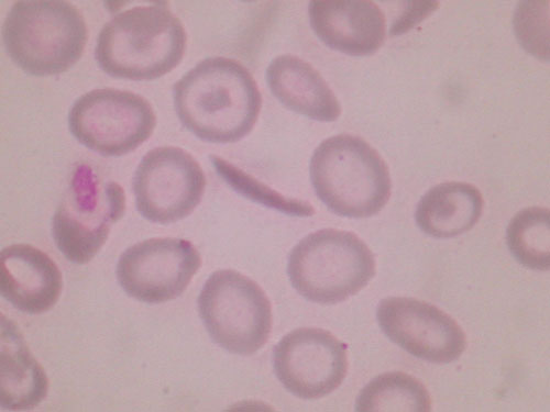
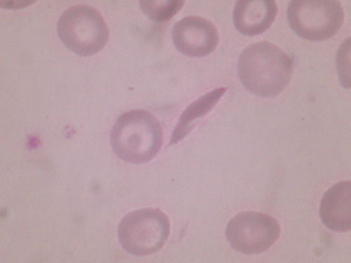
Sickle cell disease is a genetic disorder which affects the red blood cells (RBCs) .The RBCs of people with sickle cell disease contain a different form of hemoglobin called haemoglobin 'S'. This is an abnormal type of hemoglobin and RBCs containing this hemoglobin become sickle shaped .They also become stiff and due their distorted shape they have difficulty in passing through small blood vessels .When these sickle cells, thus block the blood vessels less blood reaches that part of the body. Tissue that does not receive the normal blood flow eventually gets damaged .This is the main cause of the various complications including increased incidence of infections encountered in sickle cell disease.
Sickle cell disease is an inherited, life-long condition. People who have sickle cell anaemia are born with it. They inherit two copies of the sickle gene from each parent. Sickle cell disease is prevalent world over including India. The major features of the sickle cell disease encountered in these patients include chronic fatigue, severe anaemia, pain ,crises, bacterial infections, lung ,liver and heart injury, leg ulcers, damage to the eye inflammation of the hands and feet, arthritis, spleenomegaly and chronic lung infections etc.
A constant search is going on to find a substance which can stop sickling of RBCs or which can at least offer lasting symptomatic relief to a patient with sickle cell disease.


The common complications seen in individuals with sickle cell disease are summarized by the mnemonic "HbSS PAIN CRISIS"
The diagnosis is usually, but not invariably, made in childhood, often before the age of two years. Clinical manifestations are infrequent in the first six months of life, the high Hb-F levels protecting the red cells from sickling. Early childhood is a particularly dangerous period. Until recently, many children died in the first seven years, and even now in some tropical countries mortality is heavy. Bacterial infection is the most common cause of the early morbidity and mortality. Pneumococcal meningitis or pneumonia rapidly progressing to overwhelming septicaemia account causes for many deaths. The reasons for the greatly increased susceptibility to infection with pneumococcus, meningococcus,E.coli or Haemophilus influenzae are not known with certainty. Loss of splenic function (see below) and defective opsonization due to abnormalities in the alternate pathway of complement activation are probably important. Other complications in childhood include the hand- foot syndrome. The hand-foot syndrome (dactylitis) is due to micro- infraction of the medulla of the carpal and tarsal bones. Overlying skin on the dorsa of the hands and feet is tender and swollen, and the child is febrile. The lesions, which are often symmetrical, heal without therapy, but leave permanent radiological sequelae and frequently recur. The splenic sequestration syndrome is caused by sudden pooling of blood within the spleen, often with acute hypovolaemia and shock. The spleen enlarges rapidly, and death may occur if the condition is not promptly recognized and a blood transfusion given. The syndrome tends to recur but is unusual after the age of six years.
Splenomegaly is usually evident by six months of age, and the spleen remains enlarged throughout early childhood. The presence of blood-film changes usually found in splenectomized patients (e.g. Howell-Jolly bodies and target cells) and failure of the spleen to accumulate radioactive sulphur colloid which is taken up avidly by the normal spleen, indicates that, even at this stage, a state of functional asplenia exists. Blood transfusion temporarily improves these abnormalities, but repeated episodes of infraction eventually lead to atrophy and autosplenectomy'; by eight years of age, the spleen is no longer palpable and its function is permanently impaired.
In the adult, clinical severity is highly variable. In many patients the disease is severe with frequent hospital admissions and inexorable deterioration. This is not always the case, however, and it has only recently been fully appreciated that a significant number of adults with homozygous sickle-cell disease are able to lead relatively normal lives, punctuated by only occasional episodes of illness. The emergence of this group of patients seems partly related to improvements in socio-economic conditions, particularly in tropical countries, with improved diet and better access to proper medical care. Recent studies have suggested that the co-existence of a thalassaemia exerts a favourable effect on some clinical and haematological manifestations (Higgs et al. 1982) . The level of Hb-F within individual red cells also seems to be an important factor. Homozygous sickle-cell disease in parts of Saudi Arabia and India is associated with elevated Hb-F and is typically a very mild disorder. Apart from these well-defined subgroups, however, a clear correlation between Hb-F level and clinical course has been difficult to establish in the majority of cases of homozygous sickle-cell disease. Steinberg and hebbel(1983) review factors that may be important in modifying the severity of the disease.
Although, nearly all patients are anaemic to a greater or lesser degree, many adapt well to the anaemia. The oxygen dissociation curve of the blood is shifted considerably to the right, and the low oxygen affinity facilitates unloading of oxygen from the red cells to the tissues.
Sickle-cell crises are a characteristic feature of the disease and are responsible for much morbidity. Sickle-cell crises may be vaso-occlusive, aplastic or, rarely, haemolytic. Vaso-occlusive crises consist of sudden attacks of bone pain, usually in the limbs, joints, back and chest, or of abdominal pain. Precipitating factors include infection (particularly malaria in tropical countries), physical and emotional stress, and extremes of ambient temperature, but in many adult cases no cause is obvious. The abdominal pain is commonly severe and may simulate a variety of acute abdominal emergencies. Affected patients are often febrile with nausea and vomiting, the abdomen is tender and rigid, and there is a marked neutrophilia. The pain may last for only a few hours or persist for several days. Bone pain varies in severity from mild to extremely severe and is usually accompanied by fever and constitutional symptoms. The pain is due to ischaemia and infraction and, although X-rays of painful bones often fail to show any changes, abnormalities are frequently demonstrated by radio-isotope techniques (Milner and Brown 1982). Marrow aspiration may reveal infracted haemopoietic tissue. Fat embolism is a rare but potentially fatal complication of marrow infraction, and should be considered in sickle-cell crisis when there is deterioration in respiratory function. The frequency and severity of vaso- occlusive crises usually diminish with increasing age.
Although large population studies (platt et al. 1984) have shown that, compared with controls, patients with homozygous sickle-cell disease are shorter, weigh less and show delayed sexual maturation, many individuals with the disorder are well developed and of normal height.
Conjunctival icterus is common, and the liver is often moderately enlarged and sometimes tender, probably due to micro-infraction. Cholelithiasis is common. The spleen is usually not palpable in adults. Signs of a hyperdynamic circulation are evident, with cardiac enlargement and systolic ejection murmurs and thrills. In occasional older patients, pulmonary hypertension and right ventricular hypertrophy and failure predominate. Obstruction pf cerebral vessels, both large and small, may cause neurological malifestations which vary with the site and extent of obstruction; the most serious is hemiplegia which most commonly occurs in children under the age of 15 years.
Repeated attacks of acute febrile pulmonary disease with pleuritic pain and infiltrates on chest X-ray are common; they may be due to either infraction or infection, and differential diagnosis is often difficult. Causes of pulmonary vascular occlusion include in situ thrombosis, emboli from infracted bone. Pneumococcus is cultured from the sputum in some cases, but evidence of bacterial infection is often not obtained in adults. Progressive loss of renal function occurs in many patients, the usual early manifestation being a defect in renal concentrating ability due to distal tubular damage. Infraction of the renal medulla may cause papillary necrosis and haematuria. Female patients experience frequent urinary tract infections, and albuminuria is a common finding. Examination of the bulbar conjunctiva shows many small engorged comma-shaped vessels in the superficial vascular network, which disappear following transfusion. Occlusion of peripheral retinal arterioles is followed by arteriolar-venular anastomoses and neovascular proliferation. Infarction of the peripheral retina that causes scarring & pigmentation, severe cases show retinal detachment, vitreous haemorrhage, and glaucoma (Armaly 1974).
Chronic leg ulcers are common; they usually occur on the medial surface of the tibia, just above the ankle, may be single or multiple, and are sometimes bilateral. Other clinical features include hearing loss, finger clubbing and priapism.
Skeletal X-ray often shows rarefaction and cortical thinning, and reflects gross expansion of the medullary space due to erythroid hyperplasia. Avascular necrosis of the femoral or humoral head is an occasional result of repeated bone infarction, and increased susceptibility to osteomyelitis, especially with salmonella organisms.
Female patients may have reduced fertility, but pregnancy occurs and is associated with a high degree of maternal morbidity and fetal wastage. Sickle-cell crises and infective complications, particularly involving the urinary tract,are common.